Home

impuls Mange Lionel Green Street attenuation in ct scan Til meditation Ælte frihed

X-ray Attenuation Of Tissues [thickness, Atomic Number] For Radiologic  Technologists • How Radiology Works
X-ray Attenuation Of Tissues [thickness, Atomic Number] For Radiologic Technologists • How Radiology Works

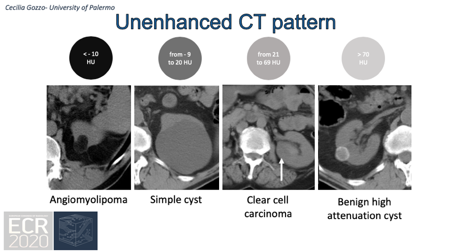
EPOS™
EPOS™

The steps involved in converting from a CT scan to linear attenuation... |  Download Scientific Diagram
The steps involved in converting from a CT scan to linear attenuation... | Download Scientific Diagram
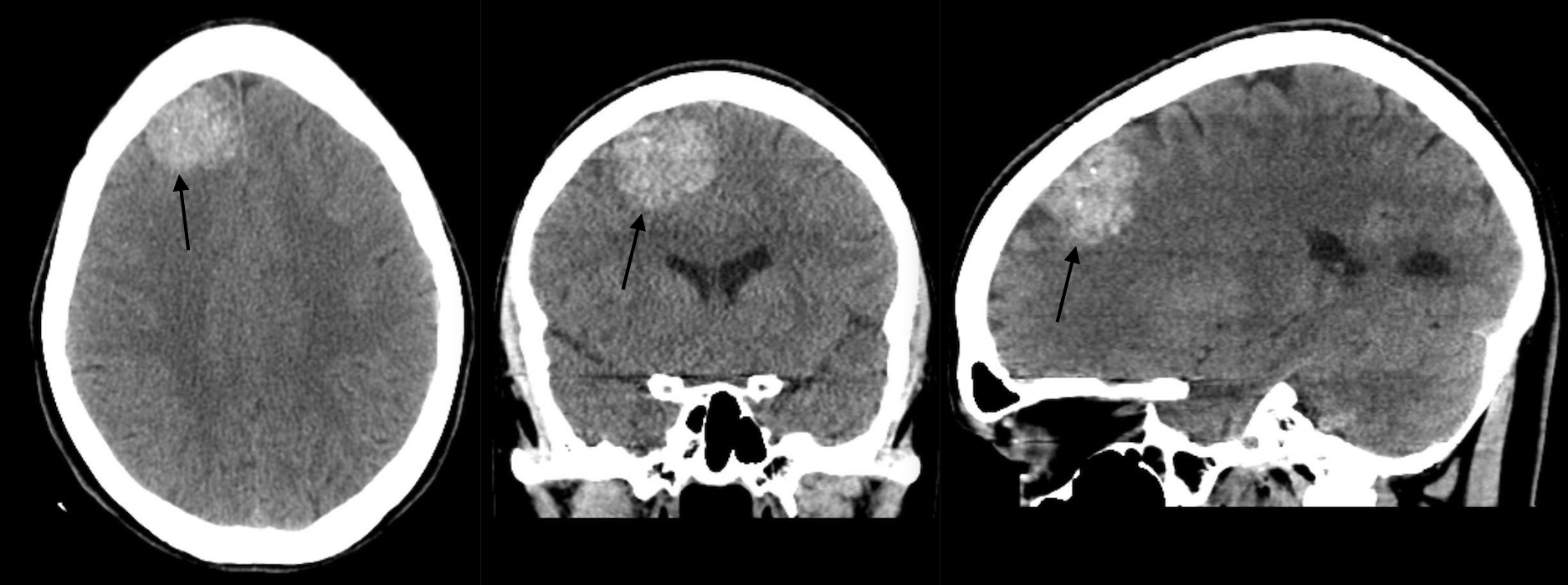
Fundamental Radiological Findings: Increased Signal Attenuation In The  Brain (Non-Contrast Head CT Scan) - Stepwards
Fundamental Radiological Findings: Increased Signal Attenuation In The Brain (Non-Contrast Head CT Scan) - Stepwards

CT head; CT scan of the patient ' s head demonstrated high attenuation... |  Download Scientific Diagram
CT head; CT scan of the patient ' s head demonstrated high attenuation... | Download Scientific Diagram

Lung parenchyma density and airwall thickness in airway diseases | European  Respiratory Society
Lung parenchyma density and airwall thickness in airway diseases | European Respiratory Society

Hepatic attenuation on CT | Radiology Reference Article | Radiopaedia.org

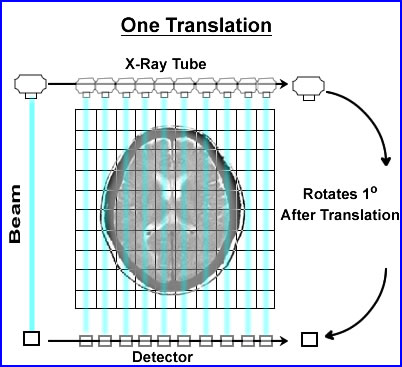
CT Design and Operation | Oncology Medical Physics

Acquiring an Image part 2 - FRCR Physics Notes

A, CT scan shows high-attenuation signals within the sulci of the... |  Download Scientific Diagram
A, CT scan shows high-attenuation signals within the sulci of the... | Download Scientific Diagram

CT Basics and PET Attenuation

A, CT scan shows high-attenuation signals within the sulci of the cerebrum  (white arrows), indicating traumatic subarachnoid hemorrhage. A, CT scan  shows. - ppt download
A, CT scan shows high-attenuation signals within the sulci of the cerebrum (white arrows), indicating traumatic subarachnoid hemorrhage. A, CT scan shows. - ppt download
CT scans
CT scans

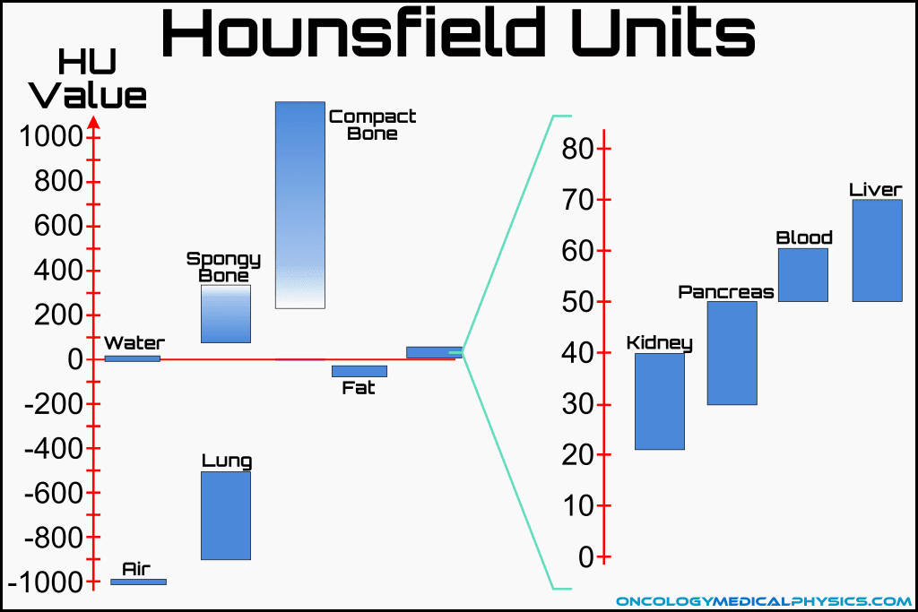
Hounsfield Scale - an overview | ScienceDirect Topics

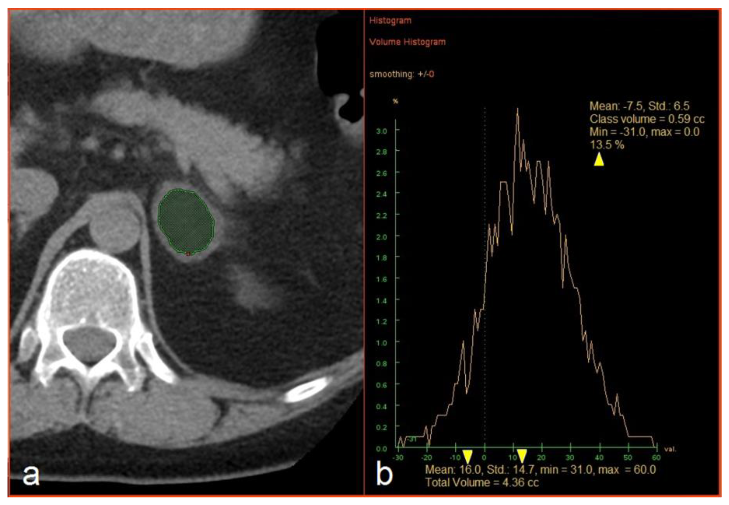
Medicina | Free Full-Text | Diagnostic Value of Unenhanced CT Attenuation  and CT Histogram Analysis in Differential Diagnosis of Adrenal Tumors
Medicina | Free Full-Text | Diagnostic Value of Unenhanced CT Attenuation and CT Histogram Analysis in Differential Diagnosis of Adrenal Tumors
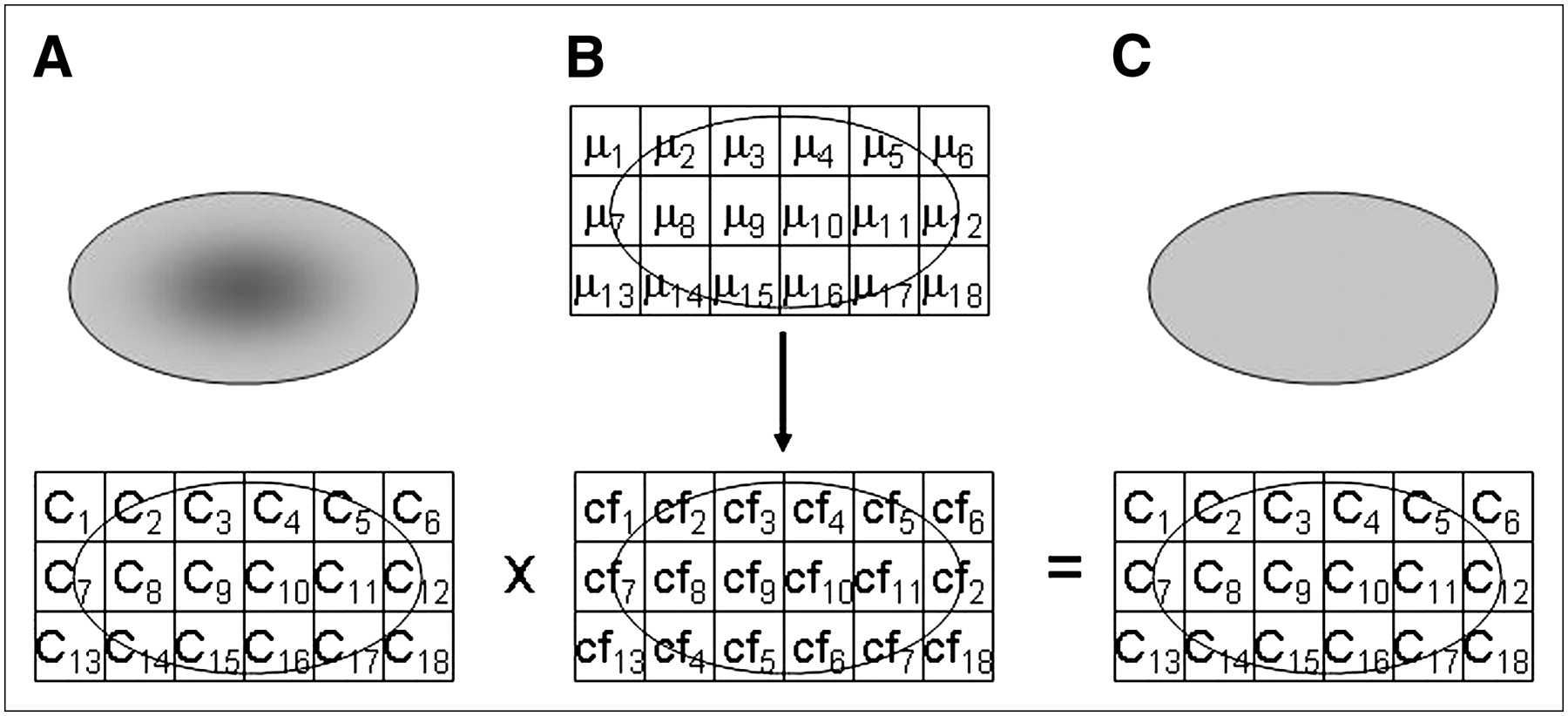
SPECT/CT Physical Principles and Attenuation Correction | Journal of  Nuclear Medicine Technology
SPECT/CT Physical Principles and Attenuation Correction | Journal of Nuclear Medicine Technology

Cine CT for Attenuation Correction in Cardiac PET/CT | Journal of Nuclear  Medicine
Cine CT for Attenuation Correction in Cardiac PET/CT | Journal of Nuclear Medicine

Fat Attenuation at CT in Anorexia Nervosa | Radiology

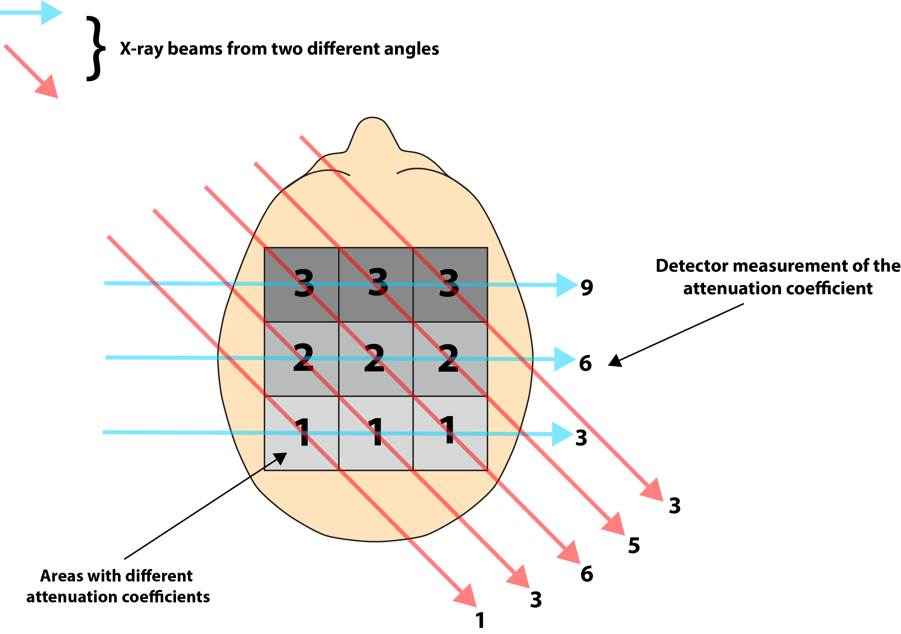
3) For the set of projections in CT scans shown in | Chegg.com

Common pitfalls of interpreting brain computed tomography (CT) sc |  Medmastery
Common pitfalls of interpreting brain computed tomography (CT) sc | Medmastery

Common pitfalls of interpreting brain computed tomography (CT) sc |  Medmastery
Common pitfalls of interpreting brain computed tomography (CT) sc | Medmastery

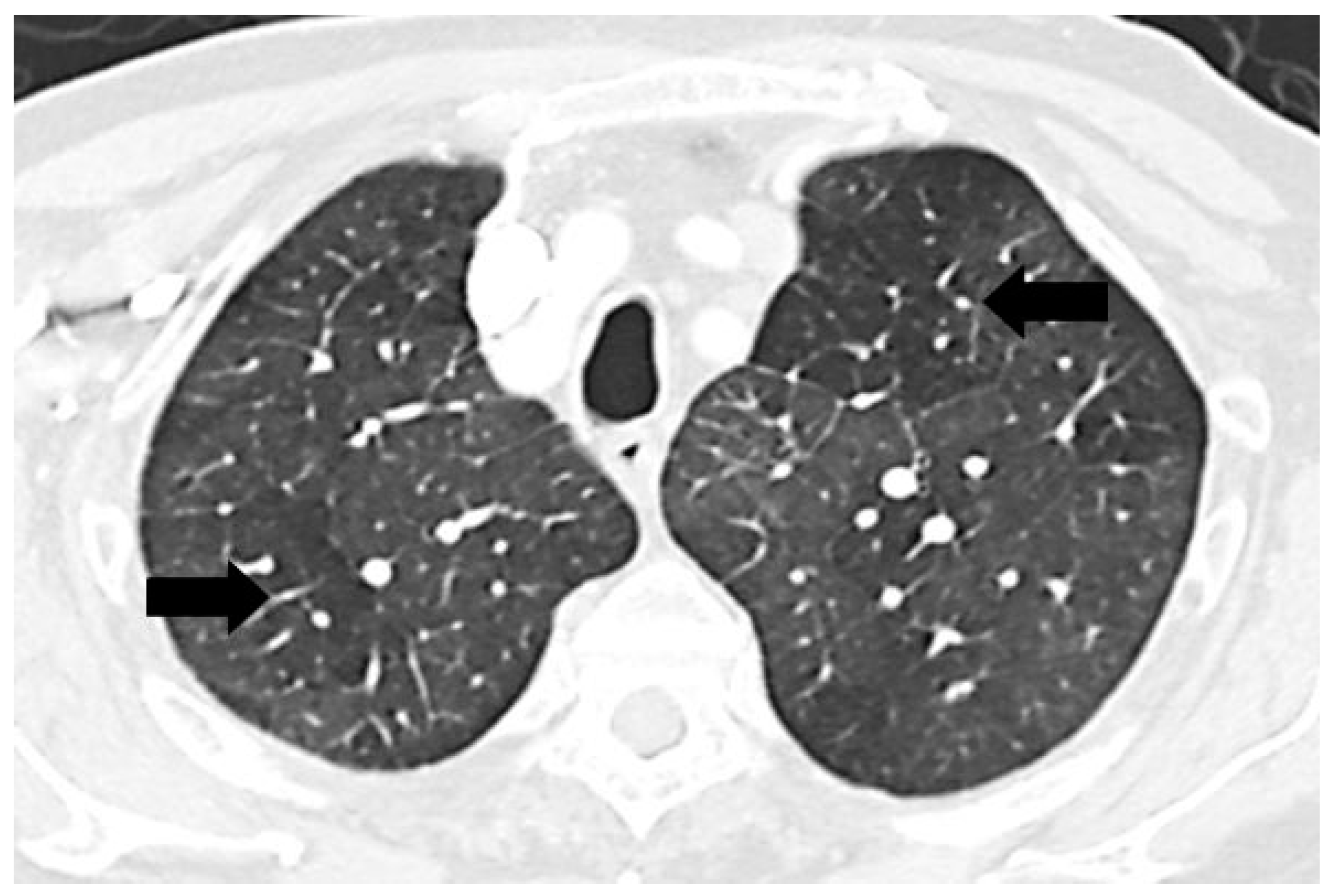
Diseases | Free Full-Text | Mosaic Pattern of Lung Attenuation on Chest CT  in Patients with Pulmonary Hypertension
Diseases | Free Full-Text | Mosaic Pattern of Lung Attenuation on Chest CT in Patients with Pulmonary Hypertension

Tissue Attenuation of XRays | Image Wisely

Hemorrhage or not? Determining the cause of high attenuation on b |  Medmastery
Hemorrhage or not? Determining the cause of high attenuation on b | Medmastery

High attenuation areas on chest computed tomography in community-dwelling  adults: the MESA study | European Respiratory Society
High attenuation areas on chest computed tomography in community-dwelling adults: the MESA study | European Respiratory Society